2. Highly sensitive “Molecular beacon” based fluorescent assay for selective detection of glutathione and cysteine
Publication:
H. Xu and M. Hepel, "Molecular beacon-based
fluorescent assay for selective detection of glutathione and cysteine",
Analytical Chemistry, 83 (2011) 813-819.
Molecular beacon based assay for GSH has been developed to address the need for screening samples with very low GSH content which is often associated with severe oxidative stress. Molecular beacons are composed of a single-stranded oligonucleotide with self-complementary 5’ and 3’ ends that can self-hybridize (Scheme 2). In the absence of a target, it forms a stem-loop structure that brings a fluorophore/quencher pair, attached to the ends of the DNA strand, into close proximity, reducing fluorescence emission. Once the single stranded loop portion of the molecular beacon hybridizes to the target, the stem melts and the resulting spatial separation of the fluorophore from the quencher leads to an enhancement in fluorescence. We have investigated a fluorescence turn-on “molecular beacon” probe for the detection of glutathione (GSH) and cysteine (Cys). The method was based on a competitive ligation of Hg2+ ions by GSH/Cys and thymine-thymine (T-T) mismatches in a DNA strand of the self-hybridizing. (Scheme 2). Scheme 2. The mechanism of turning “on” the molecular beacon by addition of GSH/Cys caused by extraction of Hg2+ ions from the MB stem and separation of the fluorophore 6-FAM (orange) from the quencher DABCYL (blue).
The molecular beacon that we have developed responds to GSH and Cys with nanomolar sensitivity (Figure 2). 5×10⁻⁹ M GSH/Cys can induce measurable fluorescence signal, indicating that the present method can successfully detect the GSH/Cys with high sensitivity. At the same condtions, very little change of the fluorescence intensity was observed upon addition of other amino acids (Figure 2d).
The outstanding ability of the molecular beacon sensing platform to respond selectively and with high sensitivity to GSH and cysteine in a matrix of amino acids is the key feature of this sensor.
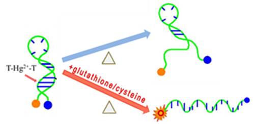
Scheme 1. The mechanism of turning “on” the molecular beacon by addition of GSH/Cys caused by extraction of Hg2+ ions from the MB stem and separation of the fluorophore 6-FAM (orange) from the quencher DABCYL (blue).
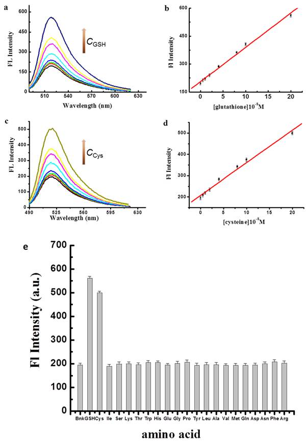
Figure 2. Fluorescence emission spectra for different concentrations of GSH (a) and Cys (c); (b,c) dependence of IFL vs (b) CGSH or (c) CCys , (e) influence of different amino acids and GSH on fluorescence emission spectrum of MB/Hg2+, [DNA] = [Hg2+] = 1×10⁻⁷ M, measurements after 15 min at 52°C; [MB] = [Hg2+] = 1×10⁻⁷ M; [amino acid] = 2×10⁻⁷ M.