7. Detection of Oxidative Stress Biomarker Homocysteine Utilizing Resonance Elastic Light Scattering
Publication: Magdalena Stobiecka, Sara Cutler, Zachary Reed, Amanda Prance, and Maria Hepel, "Detection of Oxidative Stress Biomarker Homocysteine Utilizing Resonance Elastic Light Scattering", in: Physical and Analytical Electrochemistry, S. Minteer [Ed.], ECS Transactions 28 (2010) 115-128.
Homocysteine is a biomarker of oxidative stress and a risk factor for many disorders, including cardiovascular and Alzheimer’s disease. We have investigated new methods for the development of homocysteine assays based on gold nanoparticles (AuNP) in solutions and modified solid gold piezoelectrodes. In this work, we have applied sensitive resonance elastic light scattering (RELS) and plasmonic UV-Vis spectroscopy to monitor homocysteine-induced AuNP assembly. The assembly of AuNP frameworks by covalent binding of interparticle glutaraldehyde nanobridges has been utilized for sensitive homocysteine determination. The selectivity of a glutaraldehyde crosslinking reactions toward homocysteine has been exploited to discriminate against other aminoacids which do not react with glutaraldehyde as fast as homocysteine. The measurements of a homocysteine adsorption on a solid Au electrode, carried out using the electrochemical quartz crystal nanogravimetry (EQCN), have shown that a multilayer film, with thickness of up to 3 equivalent monolayers, is formed in the zwitterionic pH range of homocysteine.
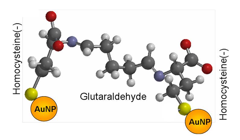
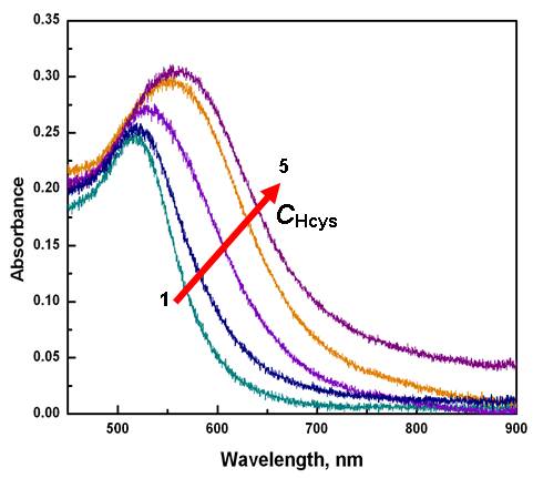
Figure 2. Absorbance spectra for glutaraldehyde linked homocysteine-capped AuNP for different concentrations of DL-homocysteine, CHcys [mM]: (1) 0, (2) 2.5, (3) 5, (4) 10, (5) 15. CAuNP = 2.53 nM, CGA = 1.12 %, pH = 7.